Breast Cancer Screening: The Age 40 vs. 45 vs. 50 Evidence Concordance
Few topics in preventive medicine have generated more public confusion than mammography starting ages. In 2009 and 2015, the USPSTF recommended routine biennial mammograms starting at age 50 (leaving ages 40 to 49 as an individual Grade C decision), while the American Cancer Society (ACS) shifted to age 45 annual screening. As analyzed by Harvard Medical School in its 2015 guideline review, both panels were looking at the same trade-off: low absolute breast cancer incidence in a woman's 40s versus higher breast tissue density that obscures 2D radiographs.
In April 2024, citing a 2% annual rise in invasive breast cancer incidence among women aged 40 to 49 and persistent mortality disparities in Black women (who face a 40% higher breast cancer mortality rate despite lower overall incidence), the USPSTF formally lowered its Grade B starting age to 40 for all average-risk women.
In April 2024, the USPSTF updated its breast cancer screening guideline from a selective Grade C to a routine Grade B for all average-risk women aged 40 to 74, recommending biennial (every 2 years) screening mammography. Starting at age 40 instead of 50 prevents approximately 1.3 to 1.5 additional breast cancer deaths per 1,000 women screened, while increasing cumulative false-positive recalls and benign biopsies.
| Screening Strategy (Digital Breast Tomosynthesis / 3D) | Breast Cancer Deaths Prevented (per 1,000) | Life-Years Gained (per 1,000) | False-Positive Recall Exams (per 1,000) | Benign Core Needle Biopsies (per 1,000) | Overdiagnosed Cases (per 1,000) |
|---|---|---|---|---|---|
| Biennial Ages 50-74 (13 total lifetime screens; 2016 USPSTF rule) |
6.7 | 121 | 850 | 140 | 12 |
| Biennial Ages 40-74 (18 total lifetime screens; 2024 USPSTF Grade B) |
8.2 (+1.5) | 165 (+44) | 1,376 (+526) | 201 (+61) | 14 (+2) |
| Annual Ages 40-74 (35 total lifetime screens; ACR / NCCN rule) |
9.9 (+3.2) | 206 (+85) | 2,193 (+1,343) | 288 (+148) | 19 (+7) |
Dense Breast Tissue: Why Mammography Sensitivity Drops and What Supplemental MRI Does
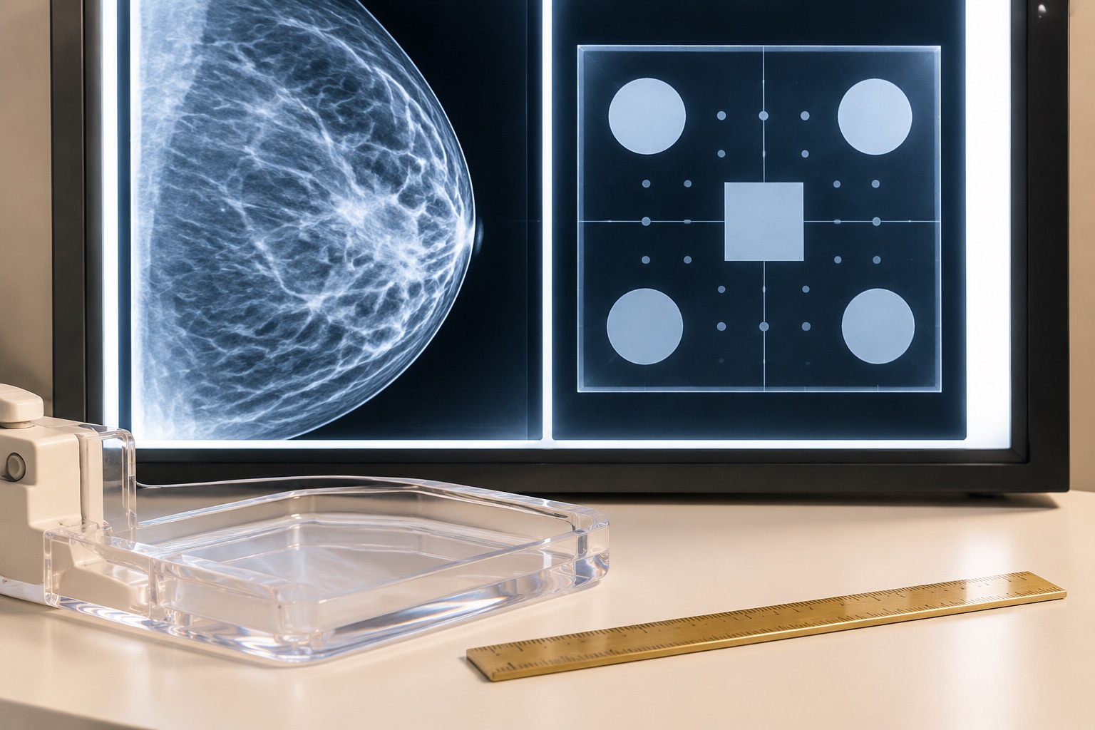
Roughly 43% of screening-age women between 40 and 74 have dense breasts: 36% have heterogeneously dense tissue (BI-RADS Category C) and 7% have extremely dense tissue (BI-RADS Category D). On a standard 2D mammogram, fibroglandular tissue and non-calcified tumors both attenuate X-rays and appear white, reducing mammographic sensitivity from ~85% in fatty breasts (Category A) down to ~50%-64% in extremely dense breasts (Category D).
Furthermore, dense breast tissue is an independent biological risk factor, increasing relative breast cancer risk 1.2-fold to 2.1-fold compared with average density. While 3D Digital Breast Tomosynthesis (DBT) acquires multiple low-dose X-ray arcs to reduce tissue overlap - raising invasive cancer detection by ~1.2 to 1.6 per 1,000 screens and cutting false-positive recalls by 15% to 20% - its sensitivity remains constrained in Category D extremely dense breasts where no adipose contrast surrounds the lesion.
Supplemental Ultrasound vs. Abbreviated Breast MRI
In the randomized Dutch DENSE trial (NEJM), women with extremely dense breasts (Category D) and a negative mammogram who underwent supplemental MRI experienced a 50% reduction in interval cancers (from 5.0 per 1,000 down to 2.5 per 1,000), detecting an additional 16.5 cancers per 1,000 first-round screens at the cost of ~80 false-positive recalls per 1,000. Whole-breast ultrasound detects ~2 to 4 additional cancers per 1,000 screens but has a substantially lower positive predictive value (PPV ~7% to 11%). Because randomized mortality data are still maturing, the USPSTF currently assigns a Grade I (insufficient evidence) to routine supplemental MRI or ultrasound in average-risk dense breasts, whereas the ACR and EUSOBI endorse supplemental MRI for Category D patients.
Frequently Asked Clinical Questions
Plain-language answers grounded in USPSTF, ACS, and 1,000-person cohort data.
Is 3D mammography (digital breast tomosynthesis) worth the radiation dose compared with 2D?
Yes, particularly when performed with synthesized 2D reconstructions (C-View). Early 3D systems required both a standard 2D exposure and a 3D sweep, nearly doubling radiation dose (though still below the FDA 3.0 mGy limit). Modern systems reconstruct the 2D image software-synthetically from the 3D tomosynthesis data, keeping the effective radiation dose (~0.4 to 0.5 mSv) virtually identical to a traditional 2D mammogram while lowering false-positive callbacks by 15% to 20%.
Should a woman with a family history of breast cancer wait until age 40 to start screening?
No. Any woman with a first-degree relative (mother, sister, daughter) diagnosed with breast cancer before age 50, or a family history of ovarian or male breast cancer, should undergo formal Tyrer-Cuzick (IBIS) or CanRisk lifetime risk assessment by age 25 to 30. Women with a calculated lifetime risk of 20% or greater qualify for annual breast MRI plus annual mammography starting at age 25 to 30 (or 10 years earlier than the youngest affected relative).